
Vaizdas:Anatomy of Human Ear with Cochlear Frequency Mapping.svg

Rinkmenos SVG peržiūros PNG dydisː 674 × 519 taškų. Kitos 5 rezoliucijos: 312 × 240 taškų | 624 × 480 taškų | 998 × 768 taškų | 1 280 × 986 taškų | 2 560 × 1 971 taškų.
Didesnės raiškos iliustracija (SVG rinkmena, formaliai 674 × 519 taškų, rinkmenos dydis: 33 KiB)
| Ši byla yra iš bendros Wikimedia Commons nemokamų resursų duomenų bazės, palaikomos Wikimedia Foundation organizacijos. Norėdami sužinoti licencijavimo smulkmenas, žiūrėkite paveikslėlio aprašymą |  |
Į paveikslėlio aprašymą |
Aprašymas
| Aprašymas |
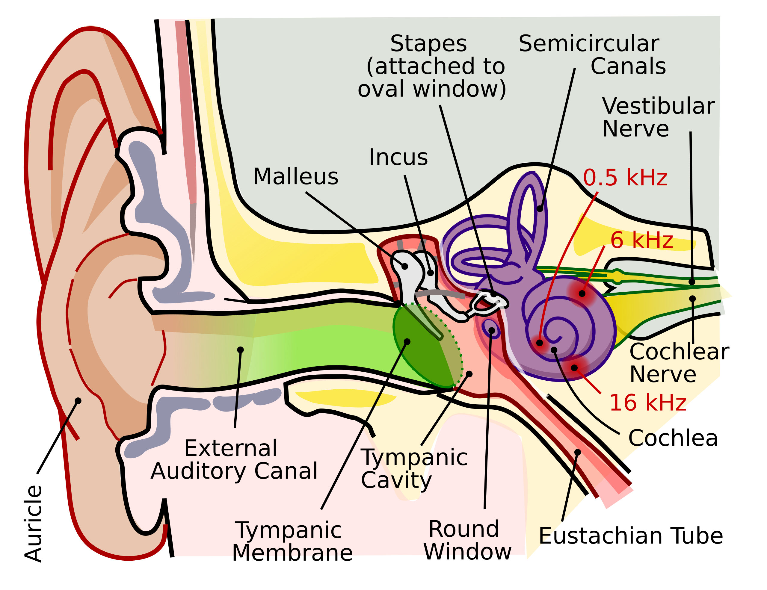
English: The human ear and frequency mapping in the cochlea. The three ossicles incus, malleus, and stapes transmit airborne vibration from the tympanic membrane to the oval window at the base of the cochlea. Because of the mechanical properties of the basilar membrane within the snail-shaped cochlea, high frequencies will produce a vibration peak near the oval window, whereas low frequencies will stimulate receptors near the apex of the cochlea (locations for three frequencies indicated schematically). Information from the cochlear receptor cells is transmitted to the cochlear nuclei via the 8th cranial nerve, and on through the midbrain to the cortex. |
| Data | |
| Šaltinis | Mano darbas (Original text: Own work by uploader, derived from File:Anatomy_of_the_Human_Ear.svg) |
| Autorius | Inductiveload |
| Leidimas (Šios rinkmenos panaudojimas kitur) |
Šiam failui taikoma Creative Commons Attribution-Share Alike 2.5 Generic licencija.
|
| Kitos versijos |
[]
|
| SVG genesis | Šis vektorinis paveikslėlis sukurtas su Inkscape. This file is translated using SVG switch elements: all translations are stored in the same file. |


Rinkmenos istorija
Paspauskite ant datos/laiko, kad pamatytumėte rinkmeną tokią, kokia ji buvo tuo metu.
| Data/Laikas | Miniatiūra | Matmenys | Naudotojas | Paaiškinimas | |
|---|---|---|---|---|---|
| dabartinis | 00:29, 17 rugsėjo 2018 | | 674 × 519 (33 KiB) | JoKalliauer | added systemLanguage="eo" |
| 20:21, 16 rugsėjo 2018 |  | 674 × 519 (32 KiB) | JoKalliauer | added systemLanguage="de" | |
| 08:33, 11 rugsėjo 2018 |  | 674 × 519 (87 KiB) | Jmarchn | Bigger (proportional real size) and full redraw (more realistic) of the auricle. Ossicles in white colour. Eardrum with contour. Added 3 labels. Add fundus to the bone and subcutaneous tissues, add superior auricular muscle, add transparency to middle ear, add separation between middle and inner ear, add division to internal auditory canal. | |
| 16:40, 29 balandžio 2009 |  | 800 × 600 (98 KiB) | Inductiveload | swap incus/malleus | |
| 18:10, 15 vasario 2009 |  | 800 × 600 (98 KiB) | Inductiveload | {{Information |Description={{en|1=The human ear and frequency mapping in the cochlea. The three ossicles incus, malleus, and stapes transmit airborne vibration from the tympanic membrane to the oval window at the base of the cochlea. Because of the mechan |
Paveikslėlio naudojimas
Paveikslėlis yra naudojamas šiuose puslapiuose:
Visuotinis rinkmenos naudojimas
Ši rinkmena naudojama šiose viki svetainėse:
- Naudojama en.wikipedia.org
- Naudojama en.wikibooks.org
- Naudojama eo.wikipedia.org
- Naudojama he.wikipedia.org
- Naudojama www.wikidata.org